Fill Out Your Anatomy Lab Heart Dissection Form
The Anatomy Lab Heart Dissection form serves as a comprehensive guide aimed at enhancing the understanding of the heart's structure and function through hands-on learning. It provides educators with crucial information about various aspects, including key anatomy, dissection techniques, and recent advancements in cardiovascular research. This form details the necessary equipment, such as sheep or lamb hearts, dissecting tools, and safety gear essential for a successful dissection experience. Spanning multiple educational stages, the document is suitable for students aged 14 to 18 and outlines learning outcomes that include identifying anatomical features, discussing heart-related diseases, and modeling surgical techniques. For teachers, helpful resources and preparatory activities are suggested to ensure students are well-prepared before engaging in the dissection. The form also highlights recent research efforts driven by the Biotechnology and Biological Sciences Research Council (BBSRC), which focus on improving heart health and addressing cardiovascular diseases through innovative treatments. By blending theoretical knowledge with practical experience, this resource aims to foster a deeper appreciation for one of the body’s most vital organs—the heart.
Anatomy Lab Heart Dissection Example
bbsrc.ac.uk
Heart Surgery
and Dissection
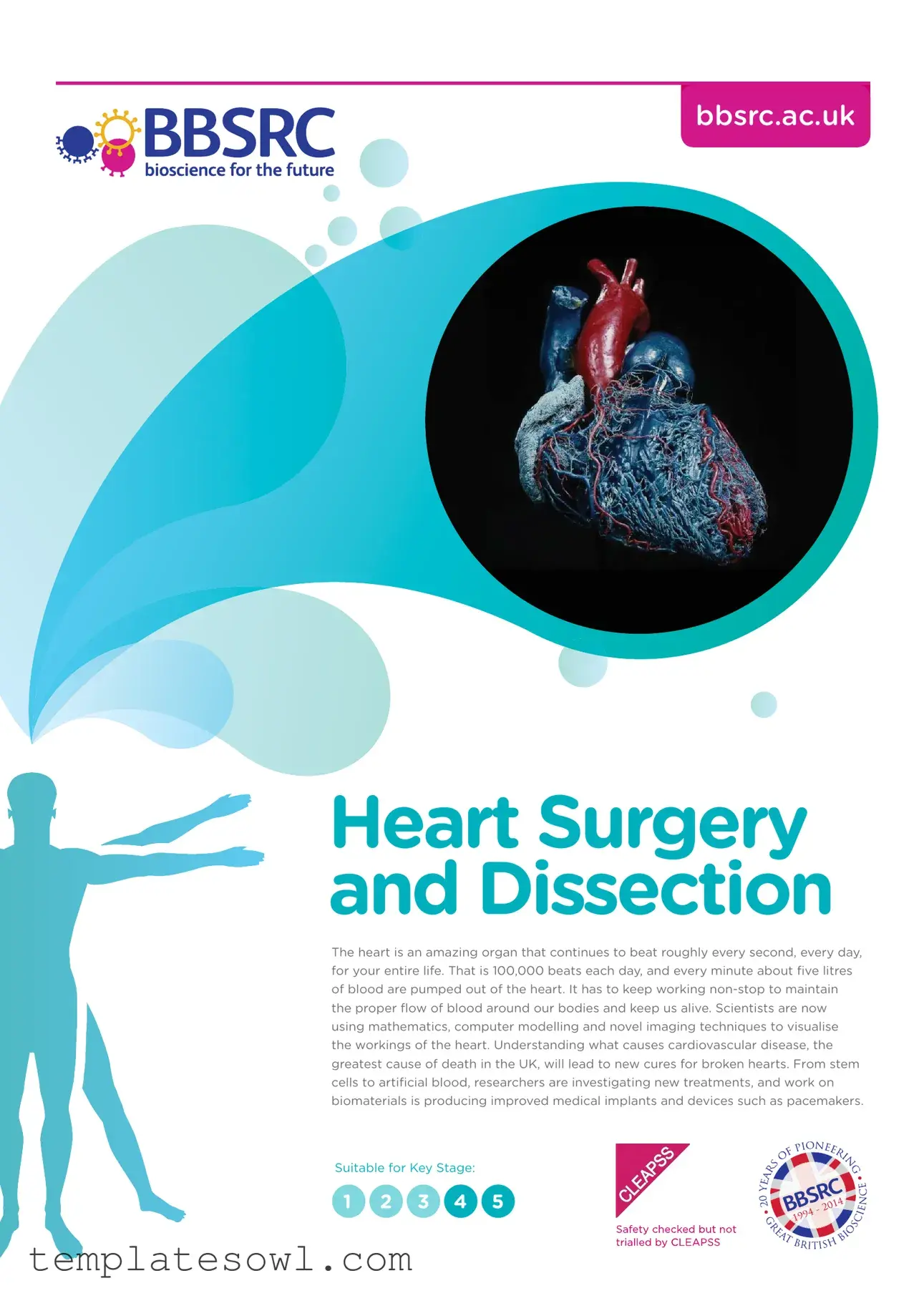
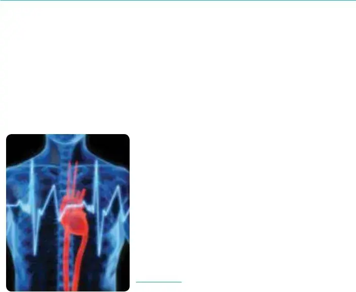
The heart is an amazing organ that continues to beat roughly every second, every day, for your entire life. That is 100,000 beats each day, and every minute about five litres of blood are pumped out of the heart. It has to keep working
Suitable for Key Stage:
1 2 3 4 5

Key Information |
Teacher |
|
Contents
02Key information
05Recent research
14Teacher preparation
15Health and safety
16Stage 1 – External anatomy
18Stage 2 – Identification and repair of heart damage
19Stage 3 – Testing blood flow through a repaired heart
20Stage 4 – Examining the internal anatomy of a heart
23Curriculum links
24Further reading
25How the heart works
29Stage 1 – External anatomy
30Stage 2 – Identification and repair of heart damage
31Stage 3 – Testing blood flow through a repaired heart
32Stage 4 – Examining the internal anatomy of a heart
33Autopsy report form
34Heart anatomy
35Card flow
36Circulation worksheet
37Missing Words
39Wordsearch
40Crossword
41Answers
44Glossary
View online
Scan the QR Code.
www.bbsrc.ac.uk |
22of4611 |
|
Key Information |
Teacher |
|
Science topics
Physiology, anatomy, cardiovascular system, exchange and transport, pathology, disease and injury
Resources
Age |
|
• |
Student sheets |
|
|
• |
PowerPoint presentation |
|
|||
|
|
|
|
|
|
|
|
Duration
125 minutes
Keywords
Heart, blood, circulation, cardiovascular, anatomy, dissection, surgery, ventricle, aorta, atrium, muscle, valve, coronary, pulmonary, artery, vein, vena cava, suture, papillary, atrioventricular, mitral, oxygen,
carbon dioxide
Learning outcomes
Students will be able to:
•Identify the internal and external anatomy of a heart
•Dissect a heart and be able to model the techniques of a heart surgeon
•Discuss heart diseases and disorders, describe how they occur, and name risk factors and possible preventative measures.
www.bbsrc.ac.uk |
32of4611 |
|

Key Information |
Teacher |
|
What you will need
•Sheep or lamb hearts
•Rubber tubing and syringe
•Dissecting equipment – trays, pins or cocktail sticks, forceps, blunt probes,
•Masking tape or stickers
•Marker pens
•Washing up bowls or access to sinks
•Curved needles
•Dental floss or fishing line
•Rulers
•Eye protection
•Waterproof aprons
•Balance
Optional
•Disposable nitrile or vinyl gloves
•Scalpels
•Slides featuring heart muscle tissue and cardiovascular pathology
•Camera for students to record the progress of their activity
Prior Learning
Students should carry out a preparatory activity to familiarise themselves with the structures of the heart. Resources such as worksheets, animations and videos can ensure students get the most from the learning session. A description of how the heart works and diagrams of the heart and circulatory system for students to label are provided.
|
Equipment |
|
|
|
|
|
© BBSRC |
|
|
|
|
www.bbsrc.ac.uk |
|
42of4611 |
|
|
|
Recent Research
The Biotechnology and Biological Sciences Research Council (BBSRC) is working towards lifelong health and
Key issues include linking changes at the molecular and cellular level to those observed at the tissue and whole organism level. A large body of evidence demonstrates that the quality and quantity of food, and dietary choice affects ageing and lifespan. There are also good data that aerobic exercise increases healthy lifespan, improves regulation of glucose metabolism and can reduce
From stem cells to artificial blood, researchers are investigating treatments for broken hearts and a variety of cardiovascular diseases. Work on biomaterials and tissue engineering is producing improved medical implants and devices such as pacemakers, as well as a range of substances with
Throughout this research, BBSRC encourages work that adopts the principles of the 3Rs (Replacement, Refinement and Reduction) in the use of animals, and aims to improve animal welfare.
Change at cellular level
© Babraham Institute

Recent Research
Can we ever mend a broken heart
Scientists at the University of Nottingham are working towards a treatment for damaged hearts. Heart disease is the most common cause of death in the UK and each year there are 20 million cases around the world. About one in five men and one in eight women die of heart disease but in the future we might be able to mend broken hearts with new heart cells.
Our hearts can fail because they are getting old, because of the stresses and strains placed on our heart by the drugs we take to treat illness such as cancer, or simply because of our genetic
Scientists are trying to develop techniques that would turn some of our own skin cells into stem cells and then turn these into beating heart cells to replace lost or damaged cells. There would be no need for drugs and we could be healed with a simple injection of our own cells.
Developing techniques
© Luchschen

Recent Research
The answer to high blood pressure may be in our brains
Blood pressure is controlled by our brains and our kidneys. Scientists are now beginning to look more closely at the brain and genes involved in the development of high blood pressure. Having high blood pressure, known as hypertension, increases the risk of stroke, heart attacks and kidney failure. You can’t ‘feel’ whether you have high blood pressure, which makes it so dangerous. Nearly one billion people around the world have hypertension.
The kidney controls blood pressure by regulating the amount of water and salt reabsorbed into the blood. High levels of salt in the blood cause the kidneys to retain water and lead to raised blood pressure. Most high blood pressure treatments target the kidneys but new research will look at ways to target the nervous system. The brain detects blood pressure using the carotid sinus, a small swelling in the carotid artery. The carotid sinus has stretch receptors that send signals to the brain when blood pressure rises. These signals go to the cardiovascular centre in the medulla and a negative feedback system sends out signals to lower heart rate and dilate blood vessels to lower the pressure.
For up to 50 per cent of patients on blood pressure tablets the treatment is ineffective and many suffer from unpleasant side effects. Researchers will explore how the genes in the brain trigger hypertension and study how ageing, exercise and a condition known as sleep apnoea affect the activity of these genes. Sleep apnoea is a blockage of airways during the night that cuts off oxygen and causes people to wake. It is associated with obesity and is often accompanied by loud snoring.
To find novel drug targets and improve current treatments, scientists will use tissue from brain banks to determine how genes are regulated in specific brain regions and how these genes interact during the development of high blood pressure. The scientists will also study how the environment may affect gene activity. In order to do this they will use a special technique that allows them to record the activity of single nerve fibres that control the diameter of arteries.

Recent Research
What causes a big heart?
Under some conditions heart cells get larger in a process known as hypertrophy. While this may sound romantic, and is necessary for developmental growth, hypertrophy often leads to heart failure. If there is a
Conditions like high blood pressure also cause the heart to grow, but in this case increased size does not improve the heart’s pumping capacity. Instead it promotes the transition to cell death and heart failure as the heart becomes prone to irregular heartbeats – arrhythmias. Researchers have discovered how signalling processes within the heart can trigger the development of enlarged heart cells which lead to heart failure. The discovery provides new insight into the mechanisms controlling cardiac growth and the processes that cause adaptation and remodelling of heart muscle. Cardiac failure accounts for 25% of deaths in the UK and is a primary cause of death in the elderly. Understanding how these pathological changes occur in the heart, in response to disease and ageing, may reveal therapeutic targets and new approaches to the treatment of heart disease.
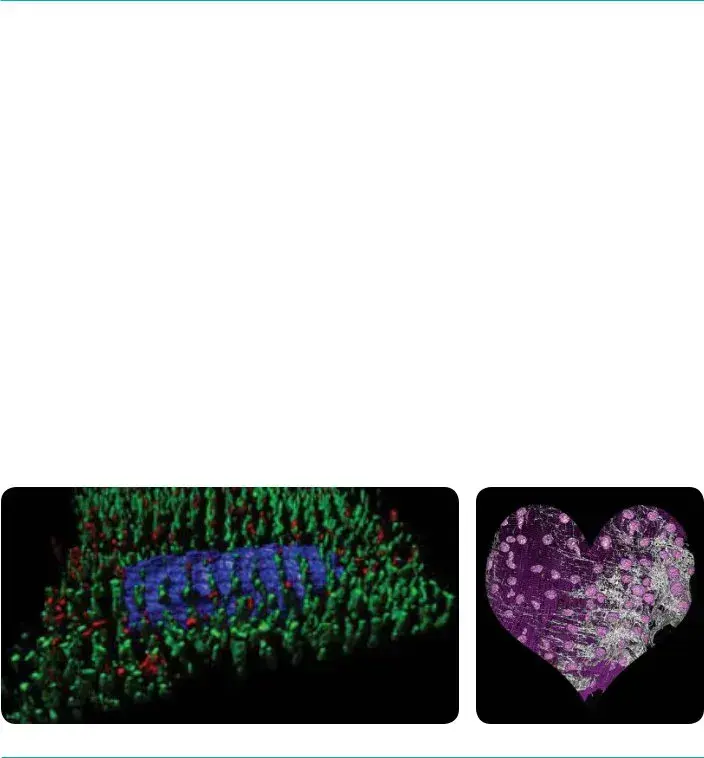
The research team found that a tiny molecule made of ribonucleic acid (RNA), microRNA, controls the levels of specific receptors produced in heart cells. MicroRNAs are copied from deoxyribonucleic acid (DNA) but do not contain code for protein. Rather, they control gene activity by binding
to specific related sequences. It is the interactions between these microRNA molecules and the receptors that promote hypertrophic remodelling of heart muscle. The receptors are channels controlling the movement of calcium ions, which are an important ‘messenger’ inside cells, regulating heart rhythm and function. Calcium ions are the link between electrical excitation of a muscle cell and its contraction. When an electrical impulse arrives at a muscle it causes calcium to enter the cell and releases calcium from internal stores resulting in contraction of the cell. If calcium signals occur at the wrong place or time, for example due to changes in receptor regulation, this can change the heart structure – decreasing its ability to pump efficiently, or triggering irregular heartbeats.
3D Reconstruction of a section through a rats heart
© Dr Llewelyn Roderick Group
Recent Research
Scientists discover the cause of a broken heart
Around
Researchers now think this ‘broken heart syndrome’ is a protective response to very high levels of adrenaline released during stress. Instead of stimulating the heart, the body responds to the adrenaline by reducing its pumping power. The same condition is sometimes seen in people who are injected with adrenaline to treat severe allergic reactions. Therefore drugs that stimulate adrenaline are likely to make the condition worse. The scientists used their animal model of the disease to investigate suitable treatments and found beneficial drugs that stimulate the heart using a different pathway to adrenaline.
Recent Research
Why some people endure exercise better than others
Exercise is essential for maintaining good health. An understanding of how elite athletes’ bodies function may help prevent elderly people developing chronic illnesses.
Researchers are studying elite athletes using
Computer models will be used to improve our understanding of why exercise tolerance is limited in sedentary or elderly individuals. The findings will be used as the basis for treating patients with heart and lung conditions who have problems with exercising.
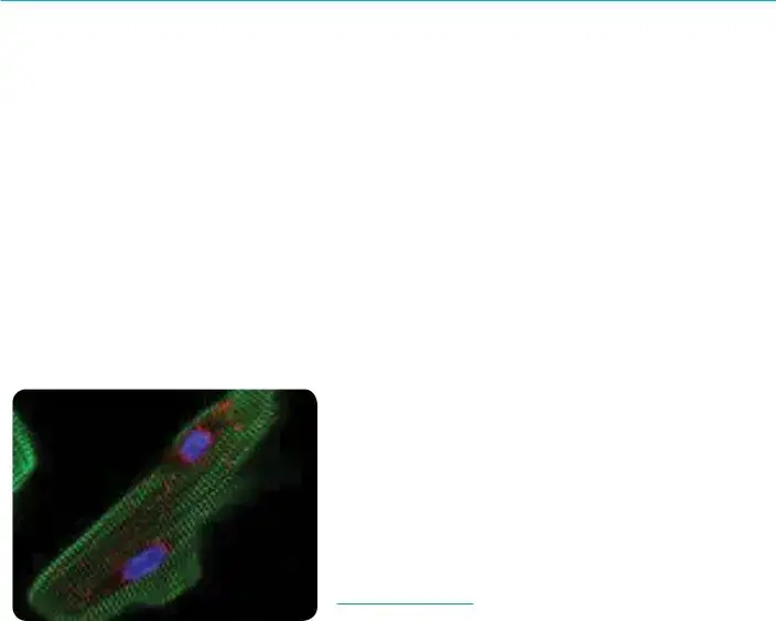
Human Heart
© Thinkstock
Form Characteristics
| Fact Name | Fact Detail |
|---|---|
| Heart Anatomy Overview | The Anatomy Lab Heart Dissection form provides a comprehensive guide to understanding both the internal and external anatomy of the heart by employing hands-on dissection techniques. |
| Learning Outcomes | Students who participate will be able to identify heart structures, perform dissections, and discuss related heart diseases and disorders, including causes and preventative measures. |
| Duration and Age Range | This instructional activity is designed for students aged 14 to 18 and is expected to take approximately 125 minutes to complete. |
| Research Backing | The content is supported by ongoing research funded by the Biotechnology and Biological Sciences Research Council (BBSRC), focusing on heart health and technologies improving cardiovascular treatments. |
Guidelines on Utilizing Anatomy Lab Heart Dissection
Completing the Anatomy Lab Heart Dissection form is a straightforward process that enhances understanding of the cardiac system. By accurately documenting observations and findings, individuals can gain deeper insights into the structure and function of the heart. Here are the necessary steps to ensure the form is filled out correctly.
- Begin by reading the form thoroughly to understand each section and what is required.
- Gather all required materials before starting. This includes your sheep or lamb heart, dissection equipment, and any safety gear.
- In the first section, carefully describe the external anatomy of the heart. Use your observations to fill in the appropriate boxes.
- Proceed to the next part, where you will identify and note any damage present in the heart. This may involve using your dissection tools for clarity.
- In the following section, outline the steps taken to repair any damage identified. Include any techniques or approaches you utilized during the repair process.
- Next, test blood flow through the repaired heart. Document your methods and observations as you conduct this test.
- Finally, examine the internal anatomy. In this section, detail your findings, ensuring to label key structures accurately.
- Before submission, review the entire form for completeness and accuracy. Make any necessary changes or additions.
What You Should Know About This Form
What is the purpose of the Anatomy Lab Heart Dissection form?
The Anatomy Lab Heart Dissection form serves to guide educators and students through the process of dissecting a heart, specifically sheep or lamb hearts. It aims to enhance understanding of the heart's structure, function, and the impact of cardiovascular diseases. By engaging in this dissection, students gain hands-on experience that complements theoretical knowledge, allowing them to visualize and appreciate the complexities of this vital organ.
What are the main learning outcomes for students participating in this dissection?
Students will come away with several key competencies. They will identify both the internal and external anatomy of a heart. Additionally, they will learn dissection techniques that mirror those of heart surgeons. The curriculum also includes discussions on various heart diseases and disorders, helping students understand how these conditions develop, their risk factors, and potential preventative measures.
What materials are required for the heart dissection?
Participants will need sheep or lamb hearts for dissection, along with various dissection tools such as trays, forceps, and scissors. Additional items like rubber tubing, a syringe, markers, and eye protection are essential for safety and effective dissection. While optional materials include gloves and scalpels, these can enhance the experience but are not strictly necessary for successful dissection.
How long does the dissection activity typically take?
The heart dissection usually lasts around 125 minutes. This duration includes time for preparation, discussion, and hands-on dissection. Adequate time is crucial for students to grasp the anatomical structures and engage thoughtfully in the activity, allowing for deeper learning and exploration.
What prior knowledge should students have before participating in the dissection?
It is beneficial for students to complete a preparatory activity before engaging in the dissection. Familiarity with the heart's structure, possibly through worksheets, animations, or videos, is crucial. Such resources will equip students with the foundational knowledge necessary for understanding the dissection process and enhance their overall engagement during the activity.
Are there safety precautions that should be considered during the dissection?
Yes, safety is paramount during any dissection. Students should wear eye protection and waterproof aprons to safeguard against potential splashes and debris. Proper handling of dissection tools is essential to prevent injuries, and instructions regarding the use of equipment should be thoroughly communicated before the dissection begins. It is also advisable to have sinks or washing-up bowls available for proper hygienic practices.
What is the significance of understanding the cardiovascular system?
Understanding the cardiovascular system is vital as it plays a crucial role in overall health. The heart is responsible for pumping blood throughout the body, delivering oxygen and nutrients to tissues, and removing waste products. Knowledge of cardiovascular anatomy and function helps students appreciate the heart's importance and the impact of various diseases, ultimately contributing to informed health decisions and preventive practices.
How do recent advancements in research relate to this dissection?
Recent advancements in heart research have highlighted the significance of understanding cardiovascular diseases, which are among the leading causes of death. The dissection activity connects students to ongoing research by illustrating the physical structure affected by these diseases. As students explore treatments like stem cells and artificial blood, they will realize the integrated nature of science and health, inspiring further inquiry into medical advancements.
What resources are available for further exploration of the heart and cardiovascular system?
Numerous resources are available to supplement learning about the heart. Students can access diagrams, videos, and academic articles that provide in-depth coverage of heart anatomy, function, and diseases. The provided curriculum links and further reading sections within the form serve as excellent starting points for both students and teachers wishing to delve deeper into these topics.
Is it necessary for students to have prior dissection experience?
No prior dissection experience is required. The Anatomy Lab Heart Dissection form is designed for both new and experienced students. For beginners, teachers can provide step-by-step instructions and support throughout the process. This encourages confidence and fosters a hands-on learning environment where all students can participate and learn at their own pace.
Common mistakes
Filling out the Anatomy Lab Heart Dissection form is an important part of any educational experience centered on understanding the heart and its functions. However, there are common mistakes that can hinder the educational process. One of the most frequent errors is neglecting to review the required resources before starting the dissection. Failing to gather necessary materials like the sheep or lamb hearts, dissecting tools, and safety equipment can lead to interruptions, detracting from a seamless learning experience.
Another common mistake involves inaccurate labeling of heart structures. It's vital to ensure that any diagrams or labels are correct for effective study later. Misidentifying parts such as the ventricle, atrium, or major arteries can lead to confusion and misunderstandings about the heart’s anatomy during discussions and examinations.
Students often overlook the significance of prior knowledge. Understanding basic heart functions and structures before dissection helps enrich the experience. Without this foundational knowledge, students may struggle to comprehend the practical aspects of the dissection, which can lead to frustration and a missed learning opportunity.
A prevalent oversight is ignoring the health and safety protocols. Not wearing the appropriate personal protective equipment, such as gloves and eye protection, not only puts students at risk but can also divert attention away from the heart dissection itself. Safety measures should always be prioritized to foster a secure and supportive learning environment.
Inadequate time management during the dissection process represents another pitfall. The Anatomy Lab Heart Dissection typically has specific durations allocated for each stage. If students rush through an initial section, they might not fully grasp essential concepts, which would affect their learning outcomes and reduce the overall value of the exercise.
Another common mistake is a lack of communication and collaboration among team members. Dissection is often best done in groups, and when students fail to discuss their observations and findings, they miss out on valuable insights that can enhance their understanding. Fostering open dialogue encourages active participation, making the process much more enriching.
Some students neglect to document their findings throughout the dissection. Keeping detailed notes or taking photos can greatly aid in the study process later. Without this documentation, recalling specific observations can become increasingly difficult, undermining the educational objectives.
Lastly, students might rush through or skip the reflection stage after the dissection. Reflecting on what was learned not only solidifies knowledge but also allows students to ponder the implications of their findings in relation to cardiovascular health. A lack of reflection can leave educational gaps and diminish the impact of the entire dissection experience.
Avoiding these mistakes contributes to a more successful and enriched dissection experience. Each step in the process is critical not only for understanding the anatomy of the heart, but also for fostering a deep appreciation of the complexities of cardiovascular health.
Documents used along the form
In the context of anatomy education, particularly during heart dissection, several documents and forms complement the Anatomy Lab Heart Dissection form. These additional resources support both the educational experience and the safe execution of the dissection procedure. Below is a list of commonly used forms and documents that typically accompany this important educational activity.
- Autopsy Report Form: This document provides a structured approach for students to document their findings and observations during the dissection. It includes sections for noting any abnormalities or detailed anatomical features.
- Heart Anatomy Worksheet: This worksheet allows students to label diagrams of the heart, reinforcing their understanding of its structure and function before hands-on dissection.
- Circulation Worksheet: This resource focuses on the circulatory system, helping students connect the anatomy of the heart with its role in blood circulation throughout the body.
- Missing Words Activity: This interactive exercise encourages students to learn vocabulary related to heart anatomy and physiology. It can enhance their understanding and retention of key concepts.
- Wordsearch: A fun tool designed to let students discover and familiarize themselves with the terminology associated with heart anatomy and dissection.
- Crossword Puzzle: Another engaging method for reinforcing vocabulary and concepts through a crossword format, encouraging critical thinking and knowledge recall.
- Answers Document: This compendium provides students with correct answers to various exercises, ensuring they can check their understanding and clarify any misconceptions.
Each of these documents plays a vital role in enhancing the hands-on learning experience in anatomy labs. By providing different ways to engage with the material, students not only gain essential knowledge of the heart’s structure but also develop critical thinking and observational skills.
Similar forms
- Autopsy Report Form: Similar in documenting anatomical findings, this form highlights external and internal examinations, helping to understand anatomical features and potential diseases.
- Medical Dissection Guide: This guide instructs students on dissecting various organs, mirroring the hands-on learning approach found in the Anatomy Lab Heart Dissection form.
- Laboratory Safety Guidelines: Both documents emphasize safety during dissections or research practices, ensuring that participants understand the dangers and necessary precautions.
- Pathology Report Template: Like the heart dissection form, this template records observations and findings related to the study of diseases affecting the heart.
- Human Anatomy Worksheet: This educational tool reinforces knowledge of heart anatomy, akin to the information presented in the dissection form.
- Heart Disease Awareness Brochure: Similar in content focus, this brochure addresses the causes and prevention of heart diseases, promoting understanding for students.
- Surgical Technique Manual: This manual contains procedures similar to the heart repair techniques outlined in the dissection form, aiding in surgical education.
- Cardiovascular System Overview: Providing an introduction to heart function and structure, this document supports the foundational knowledge required for dissection.
- Dissection Procedure Checklist: Checklist aids in systematically completing a dissection, mirroring the step-by-step stages provided in the heart dissection form.
- Research Project Proposal Template: Like the heart dissection form, this template guides the organization of research, focusing on anatomical studies or health advancements.
Dos and Don'ts
When filling out the Anatomy Lab Heart Dissection form, there are several important dos and don'ts to keep in mind. These guidelines will help ensure a smooth and effective dissection experience.
- Do read the instructions carefully before starting.
- Do use proper safety equipment, such as gloves and goggles.
- Do familiarize yourself with heart anatomy beforehand.
- Do keep your workspace organized and clean.
- Do document your findings with notes or photographs.
- Don't rush through the dissection process.
- Don't use tools that you are not comfortable handling.
- Don't forget to label any diagrams or sketches you create.
- Don't dispose of materials improperly; follow lab safety protocols.
- Don't hesitate to ask for help or clarification if needed.
Misconceptions
Here is a list of misconceptions about the Anatomy Lab Heart Dissection form:
- Dissection is only for advanced students. Many age groups can participate, including younger students. The form caters to Key Stages 1 through 5.
- You need a medical background to understand the material. The form is designed for educators and students, with clear explanations and resources that don’t require advanced knowledge.
- Only certain types of heart can be dissected. While sheep or lamb hearts are commonly used, the activities can be adapted for various educational settings.
- The dissection is purely about cutting open the heart. In addition to dissection, the form includes discussions on heart disease and how to identify its causes and impact.
- All dissection labs are the same. Each lab has specific objectives, learning outcomes, and resources tailored to enhance student understanding and engagement.
- Dissection isn’t relevant to real-world applications. Understanding heart anatomy is crucial for various fields including medicine, biology, and health science.
- It’s just about memorizing parts of the heart. The form encourages hands-on learning, critical thinking, and discussions about the functionality of the heart.
- There’s no emphasis on safety during dissection. Health and safety guidelines are carefully outlined in the form to ensure a safe learning environment for everyone involved.
Key takeaways
Filling out and using the Anatomy Lab Heart Dissection form provides essential guidance for an educational and insightful experience. Here are some key takeaways:
- Preparation is crucial. Students should familiarize themselves with the structures of the heart through resources like worksheets and videos prior to the dissection.
- Understand the learning outcomes. Students will learn to identify both the internal and external anatomy of the heart and discuss various heart diseases.
- Gather necessary equipment. A complete list includes sheep or lamb hearts, dissecting tools, and safety gear, ensuring a safe dissection environment.
- Utilize appropriate safety measures. Eye protection and waterproof aprons are essential for students during the dissection to prevent accidents.
- Document the process. Encourage students to use cameras to record their dissection steps and findings for later analysis.
- Follow the stages of dissection closely. The form outlines specific stages, from examining external anatomy to testing blood flow.
- Incorporate recent research. Discuss findings from current studies on heart health to enhance the learning experience.
- Encourage discussion. Students should actively talk about heart diseases during and after the dissection to deepen their understanding.
- Link theory to practice. Encourage students to connect the dissection with their knowledge of the cardiovascular system and how it operates.
Browse Other Templates
Erate Bear Form - Changes to filed returns should be clearly documented with an explanation when submitting an amended Form 472.
Nys Llc Dissolution - This certificate plays a crucial role in legally ending a corporation's existence.
South Carolina Sales Tax Exemption for Nonprofits - The ST-3 provides an overview of tax obligations for businesses in South Carolina.